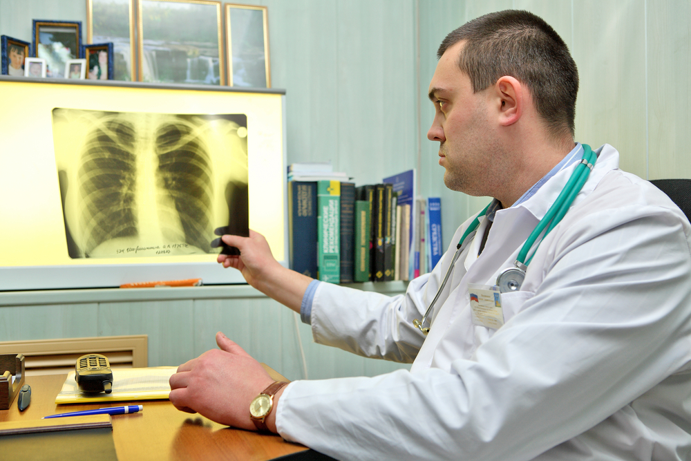
Fluorography is a method of X-ray examination, the essence of which is to photograph the tissues and organs of the human body using X-rays from a special screen with further fixation on film or digitization and displaying the resulting image on a monitor. As a rule, fluorography is used to diagnose certain lung diseases, although it was previously practiced in other branches of medicine, in particular, in gastroenterology. You will learn about who is shown the diagnosis by this method, about contraindications and the method of its implementation, as well as about what these or those changes on the fluorogram mean, from our article.
History reference
The first fluorograph was invented at the very end of the 19th century (more precisely, in 1896) by the scientist J. Blair, who is considered the discoverer of fluorography. It is interesting that for 120 years the design of the apparatus for this study has not changed radically. Of course, a number of modifications took place, but the principle of its operation remained the same as the author saw it.
At the beginning of the 20th century (in 1924), the first center for fluorographic research was opened in Rio de Janeiro, and soon this research method became widespread and spread everywhere.
The introduction of fluorography into Russian medicine was carried out by K. Pomeltsov, Ya. Shik and some other scientists. Today, every adult must undergo this study annually (starting from the age of 15), and certain categories of the population even more often (but we will talk about this below).
X-ray and fluorography - the same thing?
Indications for radiography and fluorography of the lungs are different.Despite the fact that the essence of these diagnostic methods is the same, they still differ. Fluorography is much cheaper than radiography, it involves the use of a small roll film (and the digital method does not need a film at all), which, moreover, is developed immediately by a roll, and not by each image separately.
Radiography requires the use of film of different sizes (depending on which part of the body is examined), the film is quite expensive, the images are processed individually, and special devices are needed for their development.
Therefore, fluorography is a simpler, cheaper research method, but in many cases less informative than radiography.
That is why fluorography is used as a screening (preventive) method that allows for the first time to identify or suspect a disease. If the doctor detects certain pathological changes on the fluorogram, he will recommend the patient an additional examination, among the methods of which there will be radiography.
Types, research methods
Depending on the equipment available in the medical facility, patients may be offered film or digital fluorography:
- The most common method is film. With it, X-ray radiation passes through the examined part of the patient's body (in the study of the lungs - through the chest) and enters the screen film, which is located behind it. The method provides for a fairly high (compared to digital fluorography) radiation exposure - 0.2-0.5 mSv, and the image quality is below average.
- Digital fluorography is a modern method that works on the principle of a digital camera. X-rays pass through the patient's body and fall on a special capturing matrix, after which they are digitized, and the resulting image is displayed on a computer monitor and stored in its memory. The advantages of the method are a small radiation exposure (0.05 mSv) and high quality images, which, if necessary, can be printed, sent by e-mail or saved to external media.
Indications
In our country, fluorography is an integral part of the program for the early detection of pulmonary tuberculosis. It is carried out regularly (mostly once a year) to all persons who have reached the age of 15-16. Along the way, signs of oncological diseases (in particular, lung cancer) can be detected on fluorograms.
For the purpose of diagnosing other bronchopulmonary pathologies (or, and others), this method is not used, however, their signs, of course, can be seen in the pictures.
- annual medical examination;
- living with a pregnant woman, a baby, and in severe epidemic situations - with a child of any age (certificates about the passage of fluorography by parents can be requested by preschool educational institutions or schools);
- apparatus employed;
- call for military service;
- contact with a person who is sick;
- suspicion of .
Contraindications
The main contraindications for this diagnostic method:
- children's age up to 15 years (used as a screening method for diagnosing tuberculosis in children);
- severe somatic condition (inability to be in an upright position);
- decompensated respiratory failure.
Relative contraindications are pregnancy and breastfeeding. Pregnant fluorography is prescribed according to strict indications (individual or in the case of a severe epidemic situation for tuberculosis in the region where the woman lives) and only after 25 (ideally after 36) weeks, when the organs and systems of the fetus are already formed, which means that radiation exposure will not violate them development.
It is possible to conduct this study for a woman who is breastfeeding a child, but irradiated milk should not be offered to the child - after fluorography, it must be expressed.
Preparation and methodology of the study
Fluorography of the lungs does not require any preparatory measures. The only thing is that it is advisable for the patient to stop smoking 2-3 hours before the diagnosis.
- The duration of the study is 5 minutes.
- The patient enters the fluorography room, presents the medical worker with a passport and a referral for the study.
- She strips to her waist and pulls her long hair into a high bun.
- Approaches the apparatus, stands on a special step, puts his chin in the recess available there.
- The medical worker approaches the control panel, asks the patient to take a deep breath and then hold his breath, turns on the device.
- The machine takes a picture, the patient is allowed to breathe and is asked to dress, as the procedure is over.
For the result of the study, the patient comes the next day or he is sent directly to the site - to the therapist or family doctor.
Fluoroscopy results
 If there are no pathological changes on the fluorogram, the doctor writes in the conclusion that the lungs and heart are normal.
 The description of fluorograms is carried out immediately by 2 radiologists. This is necessary in order to avoid errors in their interpretation. If there is no data on tuberculosis or lung cancer, they put a stamp on the referral and write that the lungs and heart are normal. If any changes are found in the picture, indicating the presence of a pathological process in these organs, this is reported to the local doctor or the patient himself and an additional examination is strongly recommended. In case of suspected tuberculosis, it includes:
- sputum analysis for tuberculosis (three times) - of course, if the patient has a productive cough;
Based on the results of these studies, the doctor determines the further tactics of managing the patient.
A fluorogram, in fact, like a radiograph, is an image that is formed due to the different density of the tissues through which the x-rays pass - some tissues of the rays are retained more, and others less. There is the concept of a norm, that is, this is exactly what a fluorogram of a healthy person should look like. If something does not correspond to this norm, the doctor suspects some kind of pathology:
- Most radiological changes are associated with the development of connective tissue in the lungs, which in many cases is the outcome of an inflammatory process of any nature. So, with severe bronchial asthma, a thickening of the bronchial wall will certainly be noticed.
- Cavities in the lungs, especially those containing fluid (for example, purulent masses), are usually clearly visible and look like shadows of a rounded shape with a level of fluid in them.
Local seals, such as cysts, cancer, inflammatory infiltrates or calcifications, also represent a dense tissue that retains x-rays well, not letting them through to the film - various forms of darkening are formed.
- The roots are expanded, compacted. Such a phrase in conclusion means that in the structures that form these same roots (and this is the main bronchus, pulmonary vessels - vein and artery, bronchial arteries, lymph nodes and lymphatic vessels), there is a chronic inflammatory process. Often this symptom is found in long-term smokers, while the smokers themselves may not complain. Sometimes the compaction and expansion of the roots also indicates acute inflammatory diseases, however, the patient, as a rule, has complaints, and other changes are found in the pictures, indicating in favor of a particular pathology.
- Heaviness of the roots of the lungs. Usually indicative of chronic bronchitis, is almost always found in smokers, and also occurs in people with occupational diseases, lung cancer, or bronchiectasis.
- The shadow of the mediastinum. The mediastinum is the space bounded on the left and right by the lungs (more precisely, by the pleura), in front - by the sternum, behind - by the thoracic spine and ribs. It contains such organs as the heart and aorta, trachea and esophagus, lymph nodes and blood vessels, in children - the thymus. The shadow of the mediastinum in the picture may be of normal size or be enlarged or displaced. Its expansion usually occurs with an increase in the size of the heart, and it is more often one-sided - either to the left or to the right (depending on which parts of the heart are enlarged). Its displacement is detected with an increase on one side of the pressure, which can occur with lung tumors, pneumo- or hydrothorax. This is, as a rule, a dangerous condition that requires urgent qualified medical care, therefore it is not diagnosed on fluorography.
- The lung pattern is reinforced. A pulmonary pattern is formed by the shadows of the pulmonary arteries and veins, it is visualized on any radiograph or fluorogram of the lungs. If any part of the lungs is supplied with blood more intensively than others, the pulmonary pattern on it will be strengthened. Blood flow is also activated in inflammatory diseases, as well as (tumors also use nutrients from the blood). Also, this symptom occurs in congenital and acquired, in which more blood enters the pulmonary circulation than under normal conditions. However, in such a situation, an increase in the pulmonary pattern will not be the main clinical and radiological finding. Sometimes the enhancement of the pulmonary pattern is not informative at all, but represents an error in the study - if the picture is taken not on inspiration, but on exhalation, the vessels will be filled with blood, and the vascular pattern, therefore, is enhanced.
- signs of fibrosis. The main function of fibrous tissue is the replacement of free space in the body. So, fibrosis is the outcome of a number of infectious diseases of the lungs (tuberculosis, pneumonia, and others), surgical interventions on them. In fact, it is not dangerous and speaks of a favorable resolution of the disease, but it is also a sign that part of the lung is lost, and therefore does not function.
- Foci, or focal shadows. They are shadows up to 10 mm each. This is a common and fairly informative sign, which, in combination with others, allows you to establish a diagnosis. Located in the upper parts of the lungs, focal shadows, as a rule, are signs of tuberculosis, and in the middle and / or lower parts, they indicate pneumonia. The characteristics of the foci can give the doctor an idea of the stage of the pathological process: for example, foci with uneven edges, prone to fusion, against the background of an enhanced pulmonary pattern, are a sign of the active stage of inflammation, and even edges and a high density of these shadows indicate the stage of recovery.
- Calcifications. These are rounded shadows of high (about the same as bones) density. They form when the body tries to isolate something (say, bacteria) from surrounding tissues. As a rule, Mycobacterium tuberculosis is hidden inside these calcifications, which are no longer dangerous to humans. Probably, he was in close contact with someone suffering from this pathology, received a dose of microbes from him, but good immunity did not allow the infection to develop and “buried” the microbes under calcium salts.
- Spikes. They are adhesions of the parietal and visceral sheets of the pulmonary pleura, which occur as a result of the inflammatory process. Like calcifications, they are formed then to separate the tissue affected by inflammation from healthy tissue. If the patient does not describe any subjective sensations that are unpleasant for him, adhesions are not required to be treated. If there are so many of them that they cause discomfort in a person, he cannot do without medical help.
- Pleuroapical layers. This term refers to the thickening of the pleura covering the tops of the lungs. This sign is the outcome of the inflammatory process in this part of the organ, as a rule, of a tuberculous nature.
- condition of the pleural sinuses. The sinuses of the pleura are small cavities that are located between the folds of the pleura. Their normal state is free. If liquid is found in them (otherwise - effusion) - this is a reason to be wary, since this sign indicates inflammation somewhere nearby. The sinus can be sealed, that is, there is an adhesion in its upper part - this is a consequence of a previously transferred inflammation of the pleura or another pathological process; if the patient has no complaints, it is not dangerous.
- Diaphragm. This is a large muscle that separates the chest cavity from the abdominal cavity. Its changes in the pictures can be different - flattening of the dome (one or both), its relaxation or high standing. This can be either a variant of the norm (anatomical feature), or talk about pathology (obesity, deformation by adhesions of this muscle with the pleura, a consequence of pleurisy, as well as diseases of the abdominal organs). By themselves, these signs of diagnostic value do not carry, but are always considered in combination with clinical symptoms and data from other research methods.


